


 0
0
Specifications:
| Application | Flow Cytometry | ||
| Storage Temperature | 2-8°C | ||
| Product Type | Molecular Biology Reagent | Forms | Liquid |
| Product Brand | BD Biosciences | ||
| Product Grade | Molecular Biology | ||
Preparation And Storage
Store undiluted at 4°C and protected from prolonged exposure to light. Do not freeze. The monoclonal antibody was purified from tissue culture supernatant or ascites by affinity chromatography. The antibody was conjugated with R-PE under optimum conditions, and unconjugated antibody and free PE were removed.
Recommended Assay Procedures
Cell Preparation and Staining Procedures for Conjugated Anti-Human FoxP3 Antibody
1. Bring the buffers to RT (room temperature) before use. Prepare working solutions of the BD Pharmingen Human FoxP3 Buffer Set,
Cat. No. 560098. (For Buffer A&C preparations, please see TDS of Cat. No. 560098 for details).
2. Prepare human PBMC. Dilute the cells with BD Pharmingen Stain Buffer (FBS)* to 1 X 10^7 cells/ml.
3. Pipette appropriate amount of surface staining reagent to bottom of each 12 x 75 mm tube.
4. Add 100µl of cells per tube, vortex, incubate for 20 minutes at RT protected from light.
5. Add 2 ml of wash buffer. Centrifuge 250 x g for 10 minutes, and remove wash buffer.
6. To fix cells, gently re-suspend pellet in residual volume of wash buffer and then add 2ml of 1x Human FoxP3 Buffer A. Vortex.
Incubate for 10 minutes at RT in the dark.
7. Centrifuge 500 x g for 5 minutes, and remove fixative. Caution: Be aware the pellet is buoyant.
8. To wash cells, re-suspend each pellet in 2ml of BD Pharmingen Stain Buffer (FBS)*, and centrifuge 500 x g for 5 minutes. Remove
wash buffer.
9. To permeabilize cells, gently re-suspend pellet in residual volume of wash buffer and then add 0.5 ml of 1x working solution of
Human FoxP3 Buffer C to each tube. Vortex. Incubate for 30 minutes at RT protected from light.
10. To wash cells, add 2 ml of BD Pharmingen Stain Buffer (FBS)* to each tube, centrifuge 500 x g for 5 minutes at RT. Remove buffer
and repeat wash step. Remove buffer.
11. Add conjugated FoxP3 antibody at appropriate concentrations to re-suspend the pellet. Gently shake or vortex.
12. Incubate for 30 minutes in the dark at RT.
13. Repeat wash step #10.
14. Resuspend in wash buffer and analyze immediately.
Optional: Add 300µl of 1% formaldehyde in 1x PBS and store at 4°C. Analyze cells within 24 hours.
* We recommend using the BD Pharmingen Stain Buffer (FBS; Cat No. 554656) for all wash steps and covering tubes during incubation steps with caps or parafilm. We also recommend optimizing forward scatter and side scatter voltages to visualize lymphocytes as separate from debris, red cell ghosts and/or platelets before acquisition.
** Acquire at least 15,000 to 25,000 CD4 positive lymphocytes.
Product Notices
- This reagent has been pre-diluted for use at the recommended Volume per Test. We typically use 1 × 10^6 cells in a 100-µl experimental sample (a test).
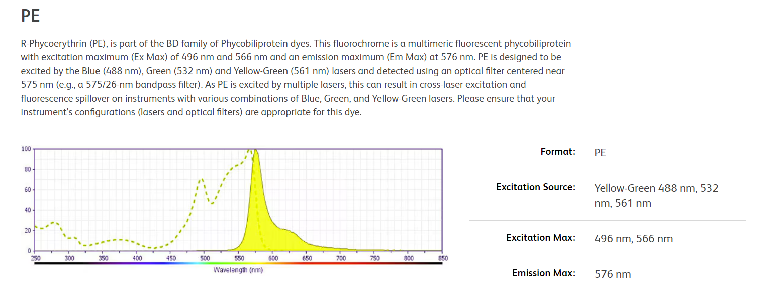
- For fluorochrome spectra and suitable instrument settings, please refer to our Multicolor Flow Cytometry web page at www.bdbiosciences.com/colors.
- Caution: Sodium azide yields highly toxic hydrazoic acid under acidic conditions. Dilute azide compounds in running water before discarding to avoid accumulation of potentially explosive deposits in plumbing.
- Source of all serum proteins is from USDA inspected abattoirs located in the United States.
- Please refer to www.bdbiosciences.com/us/s/resources for technical protocols.
- Pack Size: For 100 Tests for 25 Tests