


 0
0
Specifications:
| Application | Clinical microbiology |
| Storage Temperature | Room Temperature |
| Product Type | Test Kit |
| Product Brand | Liofilchem |
| Product Grade | Microbiology grade |
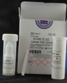
The Liofilchem® Gram Test Stick is a rapid diagnostic tool for differentiating Gram-negative microorganisms from Gram-positive microorganisms. It is designed to simplify and speed up the Gram differentiation process, enabling accurate results directly from bacterial colonies.
Key Features
- Principle: The test utilizes L-alanine-4-nitroanilide, a reagent impregnated in the stick. Upon contact with Gram-negative bacterial colonies, the reagent undergoes a chemical reaction that produces a blue-violet color when combined with the Gram Test Reagent.
- Ease of Use: Convenient stick format reduces preparation time and complexity.
- Rapid Results: Results are available within minutes.
- Applications:
- Clinical microbiology for pathogen identification.
- Routine bacterial screening in research labs.
- Quality control in industrial microbiology.
Components
- Gram Test Sticks: Pre-impregnated with L-alanine-4-nitroanilide.
- Gram Test Reagent: Used as a detector reagent to produce the colorimetric reaction.
- Quantity: The kit contains 30 sticks, sufficient for 30 tests.
Test Procedure
- Collect a Colony: Using a sterile tool, pick a colony from the bacterial culture to be tested.
- Apply to the Stick: Rub the colony onto the impregnated area of the Gram Test Stick.
- Add Gram Test Reagent: Apply the Gram Test Reagent to the area on the stick where the colony was deposited.
- Interpret Results:
- Gram-negative bacteria: Blue-violet color appears.
- Gram-positive bacteria: No color change.
Advantages
- Quick Identification: Saves time compared to traditional Gram staining methods.
- Convenient: No need for microscopes or additional equipment.
- Easy Handling: Ideal for point-of-care testing and fieldwork.
- Reliable: Produces consistent and accurate results.
Storage
- Store at 2–8°C in the original packaging.
- Protect from light and excessive moisture.
- Ensure that sticks are kept dry until use.
Precautions
- For in vitro diagnostic use only.
- Handle bacterial cultures with proper aseptic and safety protocols.
- Follow local regulations for the disposal of used sticks and reagents.
Applications
- Clinical Diagnostics:
- Differentiation of Gram-negative pathogens (e.g., E. coli, Klebsiella pneumoniae) from Gram-positive pathogens (e.g., Staphylococcus aureus).
- Research:
- Bacterial characterization and morphology studies.
- Industrial Microbiology:
- Ensures product safety by identifying contaminants in pharmaceutical and food industries.
Packaging Information
| Product | Quantity | Packaging |
|---|---|---|
| Gram Test Stick Kit | 30 sticks | Sealed in protective box |
References
- Liofilchem® technical sheet for Gram Test Stick.
- Relevant clinical microbiology guidelines for Gram differentiation.
This tool is a time-efficient alternative to conventional Gram staining, ideal for rapid diagnostics and screening in laboratory settings. Let me know if you need additional details!
Comparison: Liofilchem® Gram Test Stick vs. Liofilchem® Gram Staining Kit
| Feature | Liofilchem® Gram Test Stick | Liofilchem® Gram Staining Kit |
|---|---|---|
| Purpose | Rapid differentiation of Gram-negative from Gram-positive microorganisms. | Traditional Gram staining for detailed differentiation of Gram-positive and Gram-negative bacteria. |
| Principle | L-alanine-4-nitroanilide reacts with Gram-negative bacteria to produce a blue-violet color upon contact with Gram Test Reagent. | Crystal Violet binds with iodine to form a complex that is retained by Gram-positive bacteria but decolorized in Gram-negative bacteria. |
| Reagents Used | - L-alanine-4-nitroanilide (on the stick) - Gram Test Reagent (detector solution) | - Crystal Violet - Lugol-PVP Solution - Decolorizer Solution - Safranin Solution |
| Steps Required | 1. Apply colony to the test stick. 2. Add Gram Test Reagent. 3. Observe color change (blue-violet for Gram-negative). | 1. Fix sample on slide. 2. Stain with Crystal Violet. 3. Apply Lugol-PVP Solution. 4. Decolorize. 5. Counterstain with Safranin. 6. Observe under a microscope. |
| Results | - Gram-negative bacteria: Blue-violet. - Gram-positive bacteria: No color change. | - Gram-positive bacteria: Blue/purple. - Gram-negative bacteria: Red. |
| Time Required | Rapid (a few minutes). | Moderate (approx. 10–15 minutes). |
| Required Equipment | None. | Microscope for observation. |
| Ease of Use | Very simple and quick, suitable for fieldwork and routine checks. | Requires more steps and technical skills, suitable for detailed laboratory analyses. |
| Target Applications | - Quick screening and differentiation of bacteria. - Ideal for clinical diagnostics and quality control in industrial microbiology. | - Comprehensive bacterial analysis. - Suitable for clinical microbiology, education, and research. |
| Accuracy | Sufficient for broad differentiation but lacks detailed visualization of morphology. | High accuracy with visualization of bacterial morphology and arrangement. |
| Storage | Store at 2–8°C in original packaging. | Store reagents at 10–25°C in original packaging. |
| Shelf Life | 2 years. | 2 years. |
| Kit Components | - 30 Gram Test Sticks - Gram Test Reagent | - 250 mL each of Crystal Violet, Lugol-PVP, Decolorizer, and Safranin Solutions. |
| Training Level | Minimal training required. | Requires trained personnel for consistent results. |
| Applications | - Fieldwork - Rapid diagnostics - Screening during outbreaks | - Detailed microbiological studies - Educational demonstrations - Research investigations |
| Limitations | - Limited to broad differentiation. - Cannot determine bacterial morphology or arrangement. | - Requires a microscope. - More time-intensive compared to rapid tests. |
- Use the Gram Test Stick for rapid, point-of-care diagnostics, fieldwork, and screening applications where time efficiency is critical.
- Use the Gram Staining Kit for detailed bacterial analysis, including morphology and arrangement, in research or educational laboratories.